Introduction
The auricle can be deformed by inflammation, trauma, and neoplastic conditions. Pathologically, myxoid degeneration (MD) is a phenomenon that involves a loss of connective tissue; it affects soft tissues, including the heart valves, knee ligaments, and skin [1-5]. The pathophysiology and patient demographics of MD are poorly understood. Only one four-case series of MD of the auricle has been reported [5]. It is difficult to distinguish MD from other masses of the auricle based on a physical examination or needle aspiration due to its homogenous nature and hypertrophied cartilage lesion.
Recently, we experienced two cases of idiopathic MD of the auricle. In this case report, we present these two cases, which were managed successfully, with a literature review.
Case
Case 1
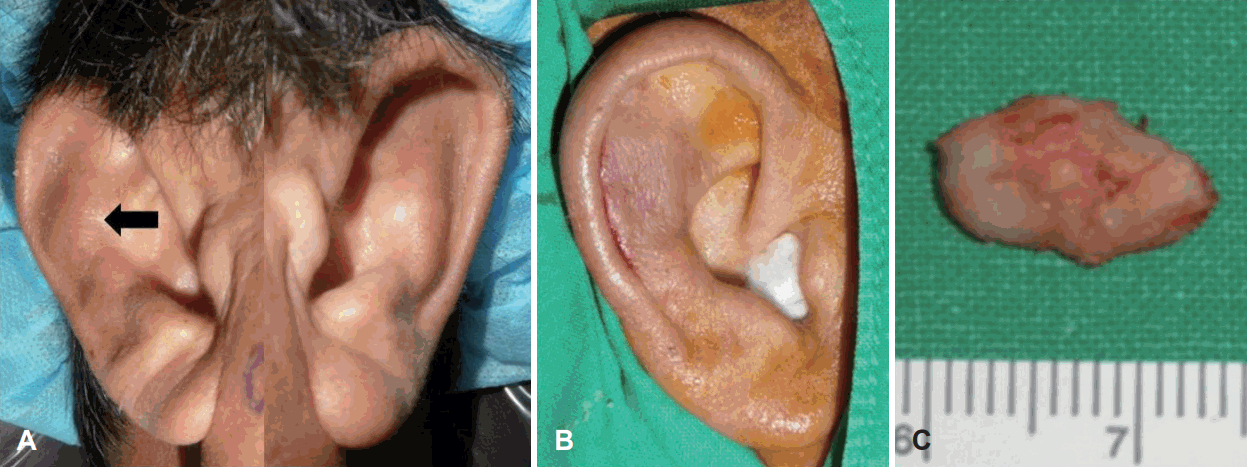
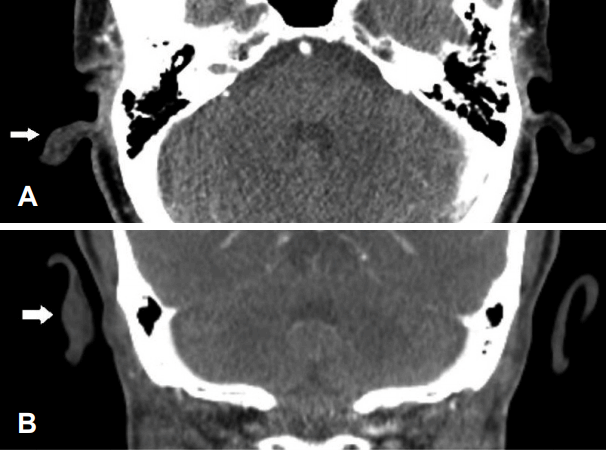
While being evaluated for Cushing’s disease and diabetes, a 51-year-old man was referred to the Department of Otolaryngology to assess a palpable lesion in his right auricle that had been present for 6 months. The lesion was primarily located in the scaphoid fossa and extended to the antihelix (Fig. 1A). On palpation, it was hard, non-tender, measured 1.6×0.8 cm, and protruded from the normal auricle without edema. The overlying skin was smooth. The external auditory canal and tympanic membrane both appeared normal by otoscopy. There was no history of disease affecting his right auricle, including trauma or tumors. He had a history of social alcohol consumption and did not smoke. Although we attempted fine needle aspiration for cytology, no tissue was aspirated. On computed tomography, the lesion had the same density as auricular cartilage (Fig. 2).
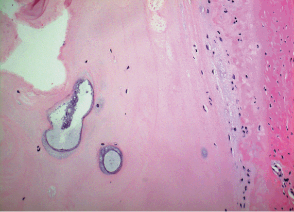
Suspecting a benign tumor, surgery was planned to obtain a pathological diagnosis and to correct the deformity for aesthetic purposes. Local anesthesia was obtained with an injection of 2% xylocaine and 1:100000 epinephrine. After making a curvilinear incision along the anterior border of the helix, the skin was elevated and the auricular cartilage was exposed. Grossly, the lesion was almost identical to the surrounding normal cartilage. The upper end of the lesion was carved away with a no. 15 blade and extended inferiorly. Finally, the 1.5√ó0.8√ó0.3 cm lesion was removed totally and the remaining auricle appeared normal (Fig. 1B and C). The wound was closed primarily with 4/0 nylon. Pathologically, the specimen showed foci of MD with loosely arranged chondrocytes compared with normal cartilage (Fig. 3). There was no noticeable nuclear pleomorphism, mitotic figures, or necrosis (hematoxylin and eosin, √ó200).
Case 2
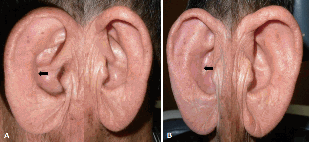
A 79-year-old man presented to the outpatient clinic of the Department of Otolaryngology with a slowly growing mass lesion in his right auricle that had been present for 3 years. The right auricle showed a non-tender, hard, 2.0√ó1.5 cm mass with a smooth surface at the medial portion of the antihelix (Fig. 4). There was no specific finding on the otolaryngologic exam and no history of illness involving his right auricle, including trauma or tumors. He had a 25-pack-year history of cigarette smoking, but did not consume alcohol. Needle aspiration for cytology failed to aspirate tissue.
An operation was performed under local anesthesia for a pathological diagnosis and to correct the deformity. After making an incision in the antihelix, surgery was performed using the same methods as in case 1. The lesion looked like normal cartilage anteriorly and a mix of cartilage and soft tissue posteriorly. It measured about 1.8√ó1.3√ó0.3 cm. The pathology was similar to that in case 1. Histological examination of the cartilage showed MD, which was similar to that in case 1 (Fig. 5).
Discussion
It is important to differentiate between benign and malignant auricle masses. Benign masses of the auricle include sebaceous cysts, keloid, hemangiomas, osteomas, and adenomas. Benign skin lesions of auricule include seborrheic keratosis, dermatosis papulosa nigra, epidermal nevus, and verruca vulgaris [6,7]. MD is distinguished from most of these diseases by its characteristic clinical manifestations and pathology.
Idiopathic MD of the auricle is extremely rare, with only four reported cases [5]. In this report, MD of the auricular cartilage was characterized by overlying normal skin, no swelling or redness around the lesions, no aspirated cells on fine needle biopsy due to the similarity of tissue density with normal cartilage and the lesion arises within the helix or antihelix and mimics normal cartilage at surgery. An important factor in the differential diagnosis of auricular MD is suspicion when a mass is associated with those characteristics. After accumulating a large number of cases of MD in the auricular cartilage with long-term follow-up results, a presumptive clinical diagnosis could be made without a biopsy.
Considering our two cases and the four in Kean and Stewart [5], MD arose on the scaphoid fossa in five cases and the antihelix in the other. Occasionally, MD has been reported in the anterior cruciate ligament (ACL) [2-4]. These patients were older, with a mean age of 43.6 years (range 35-79), and the sex distribution was unclear. Generally, they had no history of trauma. Pathologically, in the ACL, MD manifests as a homogeneous, taut, hypertrophied ACL with yellow degenerative lesions [2]. Such findings are similar to those of auricle MD in our report and in Kean and Stewart [5] We believed that aging is the only possible cause of MD in the auricular cartilage [8-10]. Age-related structural changes in the outer ear include a general loss of elasticity and muscle tonicity [11]. These structural changes in the external or middle ear may alter the external ear resonance in the elderly [12]. A large case series might help to answer this question.
Generally, MD is a benign asymptomatic condition. Kean and Stewart [5] achieved good aesthetic results by hand carving the mass via a scaphoid rim incision, which is inconspicuous and secures good exposure. We also achieved good aesthetic results without causing auricle deformity. We believe that surgical approaches should focus on confirming the pathological diagnosis with minimal aesthetic effect. This is the second report of MD of the auricle, presenting two cases. Additional studies are needed to discover the pathophysiology of MD in the auricular cartilage.